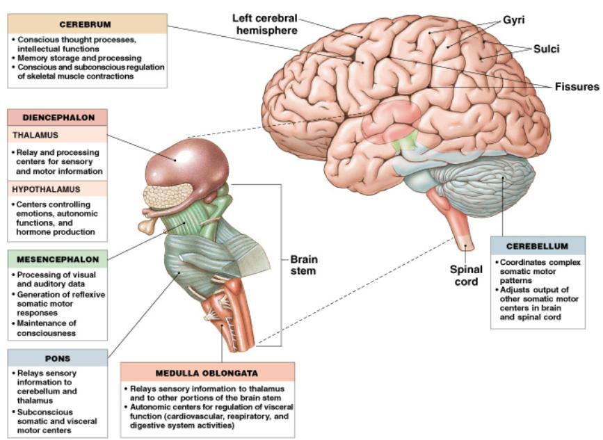
- The geography of the brain
- The brain stem
- The cerebellum
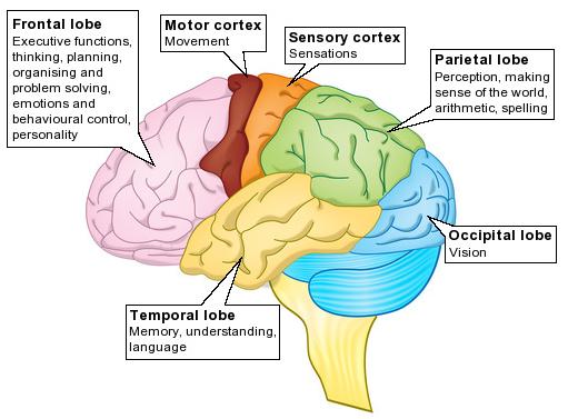
- Cerebrum
|
|
|
But First ...
| Recently, I was talking with a
cardiovascular surgeon who is doing a very new
procedure: transcatheter aortic valve replacement.
This was so interesting, and applicable to course
material, that I thought I would share it. The procedure for replacing the aortic valve is shown in the video at right.
|
Introduction to Today's Material
Last time we
talked about ...
|
|
|
 |
|
 |
A bit more on the transduction problem. Last class I mentioned that there were really 2 parts to transduction:
Here is a meter used to measure the
amount of oxygen in water.
|
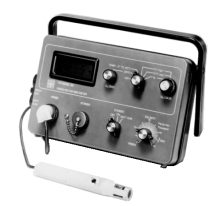 |
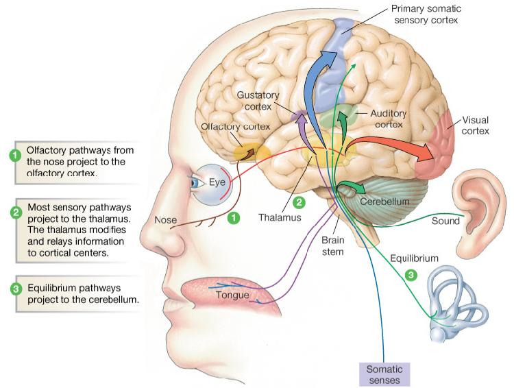
Our receptor cells are like the sensor in this example.
The circuitry in
the sense centers and association areas of our brain provide
the metering: they make sense of the current inputs.
|
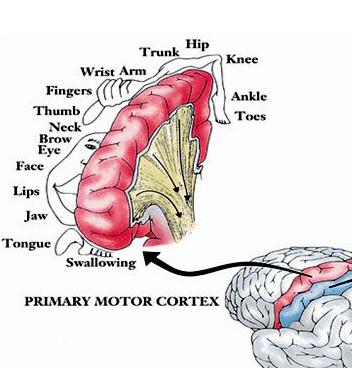 |
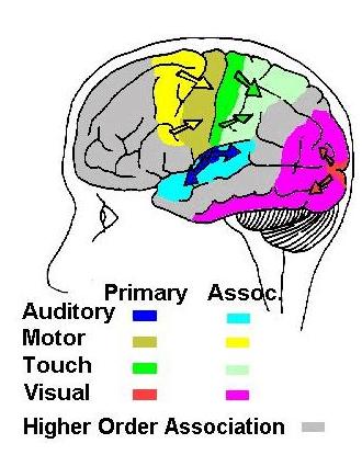
| Here is another view of the left sensory (1) and left motor (3b) cortexes. |
|
 |
Methods to Study Brain Function
Earliest Method:
Study How Brain Damage Effects Behavior and Performance
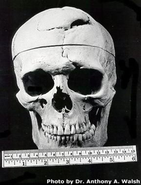
| Accidental
lobotomy of Phineas
Gage (1848)
Phineas Gage was a railroad worker in the 19th century living in Cavendish, Vermont. One of his jobs was to set off explosive charges in large rock in order to break them into smaller pieces. On one of these instances, the detonation occurred prior to his expectations, resulting in a 42 inch long, 1.2 inch wide, metal rod to be blown right up through his skull and out the top. The rod entered his skull below his left cheek bone and exited after passing through the anterior frontal lobe of his brain. |
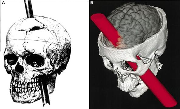 |
Remarkably,
Gage never lost consciousness, or quickly regained it (there
is still some debate), suffered little to no pain, and was
awake and alert when he reached a doctor approximately 45
minutes later. He had a normal pulse and normal vision,
and following a short period of rest, returned to work several
days later.
Effects:
|
 |
There have been
many other opportunities to study damaged brains to see how
the damage effected behavior and performance.
| Deliberately inflicted damage was the purpose of the "frontal lobotomy" (literally, removal of the frontal lobe). | 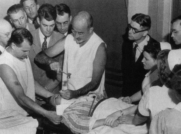 |
Injuries to the
frontal lobe (including those from lobotomies) showed multiple
personality and performance alterations.
| This table shows specific effects of frontal lobe injuries. | 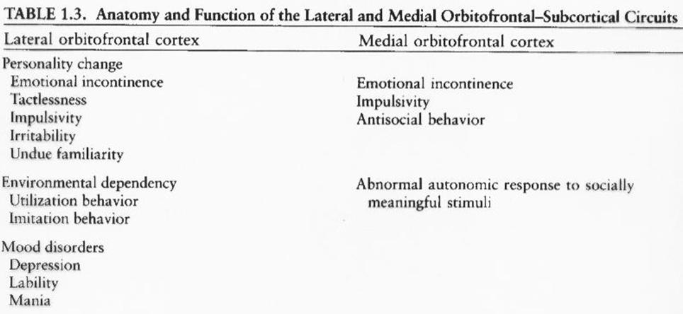 |
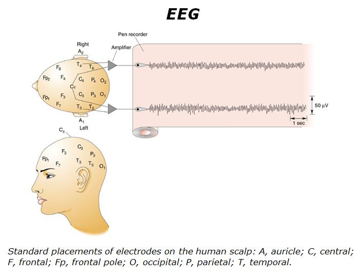
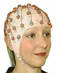
Non or Minimally Invasive Procedures
Electroencephalogram
(EEG).
|
 |
 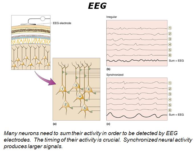 |
| The necessity for nerve activity to be synchronous to get useful information has resulted in focusing on waveforms in brain electrical activity. | 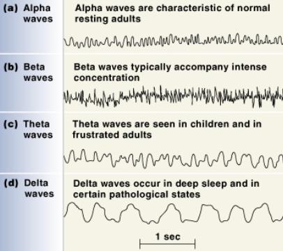 |
Positron
Emission Tomography (PET).
|
| Magnetic
Resonance Imagery (MRI).
MRI (not fMRI) is based on radio waves produced when a strong magnetic field polarizes hydrogen atoms.
|
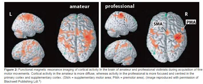
Functional
Magnetic Resonance Imagery ( fMRI ).
A fMRI
works a bit differently.
|
|
 Combined MRI / fMRI |
Some Results
from PET and fMRI Work
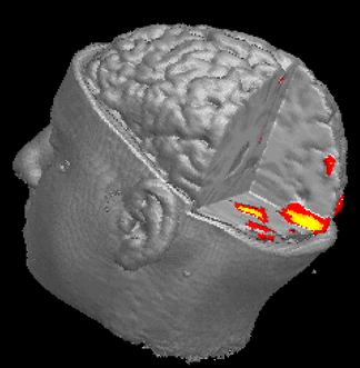
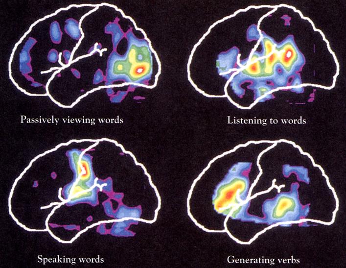
| PET and fMRI scans can show fairly clearly where specific activities occur in the brain. | 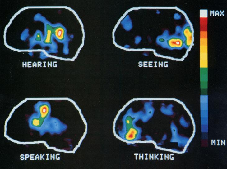 |
Pet scans
pertaining to language.
In
image to right, frontal lobe (reasoning) is left,
occipital (vision) is right, and temporal lobe
(auditory and memory) extends from right to lower
left.
|
 |
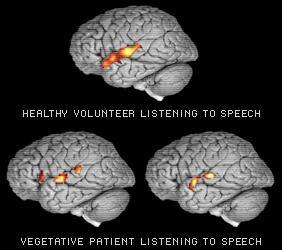
| What
does it mean to be in a "vegetative state"?
At least some of the brain centers involved with listening a functioning. |
 |
| Imagining
activity:
Subject imagined self hitting a tennis ball. What lit up? |
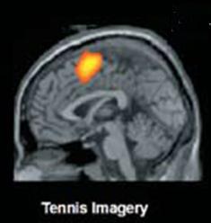 |
Second
study on a vegetative state woman.
Why
does learning hurt?
|
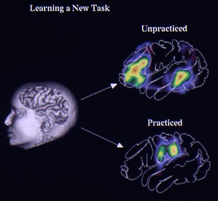 |
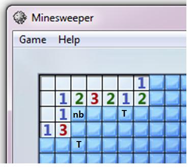
The
game Minesweeper provides an example of how the need
for thought can be replaced by pattern recognition (no
thought).
|
  |
There are many activities that we carry out without having to think much about them. In many cases, they required intense effort to learn them but, once learned, they become skills that we can apply without significant effort.
You all probably remember learning to ride a bike. Once the skill was developed, you could ride without very much thought. While learning, however, you had to work very hard.
There was a
similar huge project that you undertook that you probably
don't recall: learning to walk.
| My
grandson, Trey, began the process last summer (when he
was approaching 1 year old).
Here we see him working hard at getting up on two feet and taking a couple of steps with the support of the wall. He's got some of the neural circuitry complete, but there is more to build. (double click on the video to make it run) |
| Here we
see him taking his first two steps (several weeks
later).
He's managing all the sensory input and using it to send coordinated electrical currents to a variety of muscle groups. This is the difficult control and balancing act that we call walking. The prototype circuitry is mostly working, but needs refinement. (I don't know how to get rid of the controls in this video; anyone know how to do this?) |
| Here we
see him about a month after the last video.
He has grown the electrical circuits that allow him to be a proficient walker! Well, almost. Soon he'll walk without conciously thinking about it. |
Activities
that always require conciously using sensory input and
carefully controlling muscle output
There
are other activities that always require thought and
concentration.
|
 |
| Learning
does not happen with one reading or even a night of
cramming.
The video at right does NOT show us the way learning happens. There is no substitute for the intellectual heavy lifting that builds circuits in our brains. |
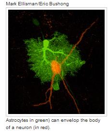
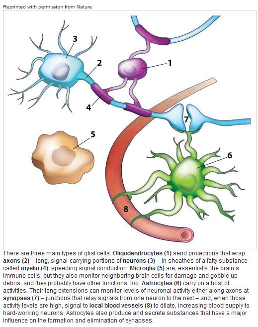
Glial Cells
In addition to
neurons, glial cells are important functional entities in the
brain. They are also very numerous (making up nearly 90% of
the brain's cells).
|
 |
It's
been known for some years that:
|
 |
Have you ever wondered why we spend so many hours every day being essentially unconscious? Surely, sleep must serve some sort of well defined physiological purpose. Although earlier studies have shown that sleep deprivation causes behavioral abnormalities, there has been surprisingly little known about the role and purpose of sleep in our lives. Here is a Ted Talk that does a nice job summarizing recent work that may answer, at least in part, why we sleep. |